Antenatal care from the first heart beat until delivery

Pregnancy Follow-up Timeline
خطة متابعة الحمل
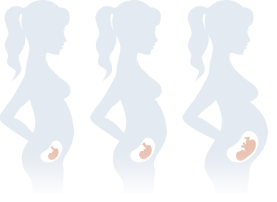
Week 4–5
At this stage, pregnancy is usually confirmed by a blood pregnancy test or an early ultrasound. The gestational sac may become visible inside the uterus, confirming that the pregnancy is developing in the correct location. Your doctor will review your medical history, estimate your due date, and start folic acid if you have not already begun taking it. Initial blood tests and urine analysis may also be requested.
في هذه المرحلة يتم عادةً تأكيد الحمل بتحليل الحمل أو بالسونار المبكر، حيث يبدأ كيس الحمل في الظهور داخل الرحم، مما يؤكد أن الحمل موجود في مكانه الصحيح. كما يتم مراجعة التاريخ المرضي، وتحديد موعد الولادة المتوقع، والبدء في تناول حمض الفوليك إذا لم يكن قد بدأ من قبل، مع طلب التحاليل الأساسية وتحليل البول عند الحاجة.
Week 6–7
Around six to seven weeks, the baby’s heartbeat is usually visible on ultrasound. The fetal pole and crown-rump length (CRL) are measured to accurately determine the gestational age. If everything appears normal, your pregnancy follow-up plan will be discussed, together with nutrition, lifestyle advice, and the supplements you should continue taking.
في الأسبوع السادس إلى السابع يظهر نبض الجنين في أغلب حالات الحمل، ويتم قياس طول الجنين (CRL) لتحديد عمر الحمل بدقة، وهو أدق قياس لتحديد موعد الولادة المتوقع. كما يتم وضع خطة متابعة الحمل، ومناقشة التغذية السليمة، والفيتامينات والمكملات الغذائية المناسبة خلال هذه المرحلة.
Week 9
By nine weeks, all the baby’s major organs have started to form. The ultrasound confirms normal fetal growth and a regular heartbeat. Most mothers continue folic acid until the end of the first trimester, while additional supplements such as vitamin D or progesterone are prescribed only when medically indicated.
بحلول الأسبوع التاسع تكون معظم أعضاء الجنين الأساسية قد بدأت في التكوين، ويُستخدم السونار للتأكد من نمو الجنين بصورة طبيعية واستمرار نبضه بصورة منتظمة. يستمر تناول حمض الفوليك حتى نهاية الثلث الأول من الحمل، بينما تُستخدم المكملات الأخرى أو البروجستيرون فقط إذا أوصى بها الطبيب.
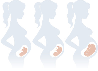
Weeks 11–13+6
This is one of the most important ultrasound appointments during pregnancy. A detailed first-trimester scan is performed to measure the Nuchal Translucency (NT), assess the presence of the nasal bone, measure the crown-rump length, evaluate the baby’s early anatomy, and determine the risk of certain chromosomal abnormalities when combined with blood tests or non-invasive prenatal testing (NIPT), if indicated.
تُعد هذه من أهم زيارات الحمل، حيث يتم إجراء السونار التفصيلي للثلث الأول من الحمل. يتم قياس سمك القناة العنقية (NT)، والتأكد من وجود عظمة الأنف، وقياس طول الجنين، وفحص تكوين الأعضاء بصورة مبدئية، بالإضافة إلى تقييم احتمالية بعض اضطرابات الكروموسومات عند دمج نتائج السونار مع تحاليل الدم أو اختبار NIPT إذا كانت هناك حاجة لذلك.
Weeks 18–22
Between 18 and 22 weeks, the detailed fetal anatomy scan is performed. This examination carefully evaluates the baby’s brain, face, spine, heart, lungs, diaphragm, stomach, kidneys, bladder, limbs, umbilical cord, placenta, amniotic fluid, and cervix. It is also the best time to detect most structural abnormalities and assess your baby’s overall development.
بين الأسبوع الثامن عشر والثاني والعشرين يتم إجراء السونار التفصيلي، وهو أهم فحص بالموجات فوق الصوتية خلال الحمل. يتم خلاله تقييم جميع أعضاء الجنين بالتفصيل، بما في ذلك المخ، والوجه، والعمود الفقري، والقلب، والرئتين، والحجاب الحاجز، والمعدة، والكليتين، والمثانة، والأطراف، والحبل السري، والمشيمة، وكمية السائل الأمنيوسي، بالإضافة إلى قياس طول عنق الرحم. ويُعتبر هذا الفحص أفضل فرصة لاكتشاف معظم العيوب الخلقية ومتابعة نمو الجنين بصورة دقيقة.
Weeks 27–28
At this stage, screening for gestational diabetes is usually performed. A complete blood count may be repeated to assess for anemia, and iron supplementation is commonly started or adjusted if needed. Calcium supplementation is also recommended for most pregnant women, while blood pressure and fetal growth continue to be monitored.
في هذه المرحلة يتم غالبًا إجراء اختبار سكر الحمل، وإعادة صورة الدم لاكتشاف الأنيميا إذا وُجدت، كما يبدأ تناول الحديد أو يتم تعديل جرعته حسب نتائج التحاليل، ويُنصح أيضًا بالاستمرار على الكالسيوم وفقًا لتعليمات الطبيب. كذلك تتم متابعة ضغط الدم ونمو الجنين بصورة منتظمة.
Weeks 29–31
A growth ultrasound is usually performed to evaluate your baby’s weight, growth pattern, amniotic fluid volume, placental function, and fetal movements. Doppler studies may be added when medically indicated to assess blood flow between the placenta and the baby.
يتم خلال هذه الفترة إجراء سونار لمتابعة نمو الجنين، وتقدير وزنه، ومعدل نموه، وكمية السائل الأمنيوسي، ووظيفة المشيمة، بالإضافة إلى تقييم حركة الجنين. وقد يتم إجراء فحص دوبلر عند الحاجة للاطمئنان على تدفق الدم بين المشيمة والجنين.
Weeks 36–37
As delivery approaches, your baby’s position is confirmed to determine whether the head is down. A final ultrasound assesses fetal growth, placental location, and amniotic fluid. Group B Streptococcus (GBS) screening may be performed according to local guidelines. This visit is also the ideal time to discuss your birth plan, including vaginal delivery, cesarean section, and available pain relief options such as epidural analgesia.
مع اقتراب موعد الولادة يتم التأكد من وضعية الجنين، وما إذا كان الرأس قد استقر في الحوض. كما يُجرى سونار أخير لتقييم نمو الجنين، ووضع المشيمة، وكمية السائل الأمنيوسي. وقد يتم إجراء مسحة بكتيريا المجموعة العقدية (GBS) وفقًا للبروتوكولات المتبعة. وتُناقش خلال هذه الزيارة خطة الولادة، سواء كانت ولادة طبيعية أو قيصرية، بالإضافة إلى وسائل تخفيف الألم مثل التخدير فوق الجافية (إبرة الظهر).
Weeks 39–40
At full term, your doctor will monitor your baby’s well-being with ultrasound and fetal heart monitoring whenever indicated. If labor has not started naturally, the cervix will be assessed and induction of labor may be discussed when medically appropriate. During labor, epidural analgesia is available for pain relief during vaginal birth, while spinal anesthesia is the most common and preferred anesthetic technique for planned cesarean delivery.
عند اكتمال الحمل تتم متابعة صحة الجنين بالسونار ومراقبة نبضه عند الحاجة، وإذا لم تبدأ الولادة تلقائيًا يقوم الطبيب بتقييم عنق الرحم ومناقشة تحفيز الولادة إذا كان ذلك مناسبًا طبيًا. أثناء الولادة الطبيعية يمكن استخدام التخدير فوق الجافية (إبرة الظهر) لتخفيف الألم، بينما يُعد التخدير النصفي هو الخيار الأكثر شيوعًا وأمانًا في الولادة القيصرية المخطط لها.